Intracellular molecular dynamic analysis

- researcher's name
- affiliation
- keyword
-
background
The higher brain functions of memory and learning are supported by a phenomenon known as neuron synapse plasticity. Detailed clarification of the operating principle of this cell signal transfer mechanism will lead to elucidation of brain function and application to the development of drugs for treating abnormalities.
summary
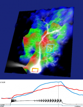
Using random scan two-photon excitation microscopy, we can measure the movement of intracellular molecules. In particular, the calcium ions (Ca2+) and phosphoenzymes, receptors and other proteins involved in the synapse plasticity of neurons are the target for this analysis.
application/development
●Development of image acquisition programs for two-photon excitation microscopy.
●Searching, visualization and dynamics analysis of target proteins in drug development (molecules that activate or inhibit receptors and enzymes involved in synapse plasticity of neurons) using the high speed of two-photon excitation microscopy.
predominance
By introducing a random scanning system based on an AOD (acousto-optic deflector) to the technology possessed by our laboratory, we can observe the scattering of intracellular free proteins (formerly undetectable) at multiple points, at a high speed of maximum 100kHz. Moreover, the damage to the cell is lower than previous measurement methods and it is possible to obtain a three-dimensional image of deep tissue. FCS (fluorescence correlation spectroscopy) recording that uses this high speed is a uniquely developed analytical method, through the use of which, it is possible to observe the behavior of different proteins at various sites in the cell.
purpose of providing seeds
Sponsord research, Collaboration research, Technical consultation
material
posted:
2014/05/21


